Publié le
Lecture 8 mins
New 3D embryo-foetal imaging using immunostaining
Presented by Gérard COULY, Work from the Institut de la Vision [Vision Institute], Inserm UMRS 968, Paris
Clarifying human development using 3D imaging of transparent whole embryos and organs
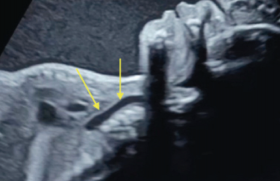
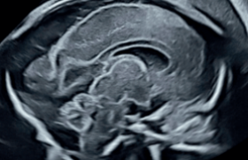
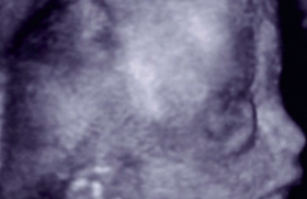
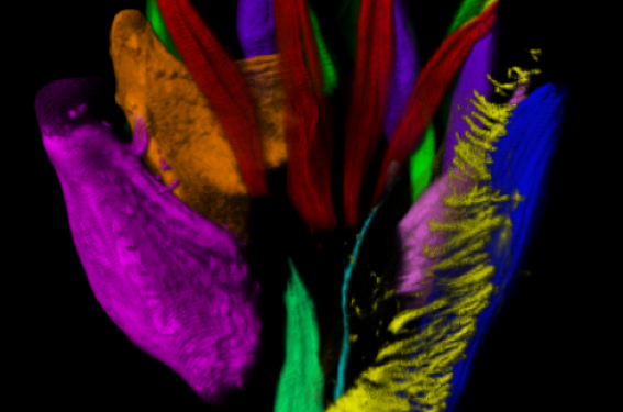
Generating a precise cellular and molecular cartography of the human embryo is essential to understand the mechanisms of organogenesis in normal and pathological conditions. Here, we combined whole-mount immunostaining, 3DISCO clearing and light-sheet imaging to start building a three-dimensional cellular map of human development during the first trimester of gestation. We provide 3D images at an unprecedented resolution of the developing peripheral nervous system, vascular system, cardiopulmonary system, urogenital system and muscular system. We show that the adult-like pattern of skin innervation is established before the end of the first trimester with important intra- and inter-individual variations in nerve branches. We also present evidence for a differential vascularization of male and female genital tracts concomitant to sex determination. This work paves the way for a cellular and molecular reference atlas of human cells, which is critical to understanding human developmental disorders.
Introduction The Institut de la Vision work group, led by Alain Chedotal (UCPM, Inserm UMRS 968, Paris 6 Sorbonne), published the imaging of numerous developing organ structures from embryos and foetuses, shown using immunostaining, from the end of the first trimester to the beginning of the second...
Attention, pour des raisons réglementaires ce site est réservé aux professionnels de santé.
pour voir la suite, inscrivez-vous gratuitement.
Si vous êtes déjà inscrit,
connectez vous :
Si vous n'êtes pas encore inscrit au site,
inscrivez-vous gratuitement :